A 22-year-old bridge, retired with dignity — Ms. Abida's full-mouth rebuild.
Ms. Abida Rashid is a 43-year-old Lahore patient who came to our Engineers Town clinic carrying an extraordinary piece of dental history — a full-mouth fixed bridge that had been placed when she was in tenth grade, more than two decades earlier. The bridge had served her loyally. It had also slowly run out of margin. This is the full story of the staged endodontic, periodontal and prosthetic rehabilitation that gave her a new, properly maintained mouth.
 Before
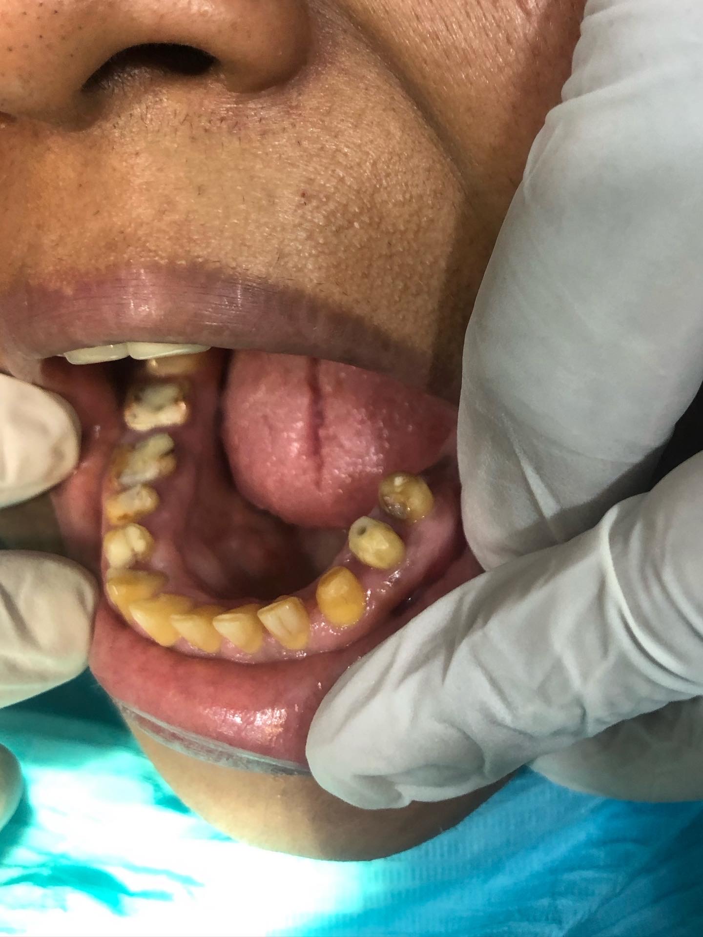 After
AfterA bridge from a different era. A patient who needed a careful exit plan.
The hardest part of a long-overdue rehabilitation is rarely the technical work. It is the careful sequencing — what comes out, what comes off, what gets treated underneath, and when. Ms. Abida's case is a good example of how an honest plan beats a fast one.
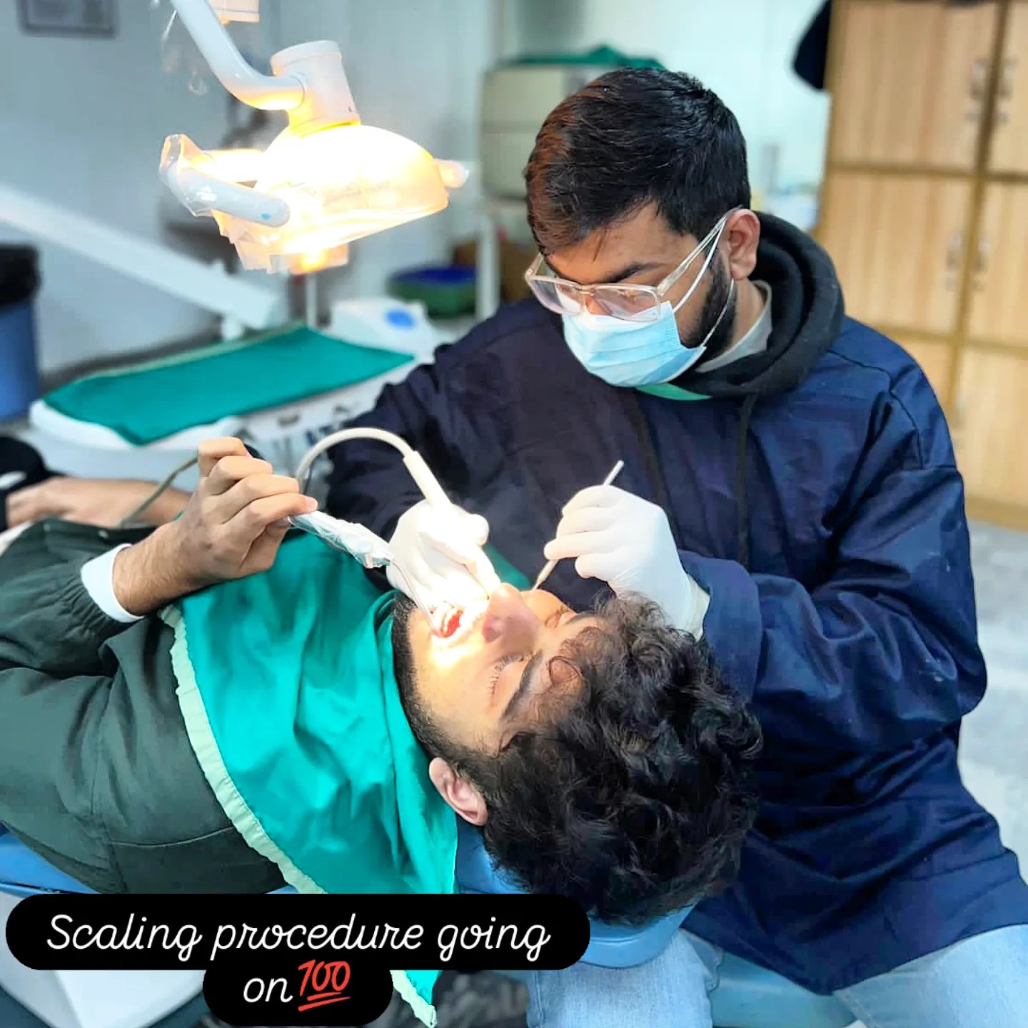
Ms. Abida Rashid came to our clinic in mid-2024 with a single, slightly understated complaint — “there's something not right under the bridge.” She is 43 years old, the mother of two teenagers, and a long-time resident of the same neighbourhood as our clinic. She had not seen a dentist for routine maintenance in roughly six years. The bridge in her mouth was, by her own account, the result of treatment done when she was in tenth grade — placed by a dentist who had since retired and whose records were no longer accessible.
That meant we were starting from a clean slate. We had a 43-year-old patient with a 22-year-old prosthesis that she could no longer rely on, a vague description of “something not right”, and no prior charting to compare against. We did what we always do in this situation — we treated her first appointment as a full diagnostic visit and explained, before we did anything, that we would almost certainly not be doing any treatment that day.
The clinical examination took 45 minutes. We charted every tooth that was either visibly part of the bridge or part of the natural dentition. We took two periapical X-rays of each quadrant, plus a single panoramic radiograph, plus six intra-oral photographs. We probed the gum at six points around every bridge abutment, and we tested cold-stimulus response on every accessible tooth surface.
What we found could be summarised in four sentences. The bridge had been placed over multiple teeth without root canal therapy where the depth of preparation should have made that necessary.Several of those teeth had developed silent pulpal pathology over the intervening two decades.The gum at the margins of the bridge was visibly hyperplastic, with multiple sites of bleeding on probing. The alveolar bone supporting the abutment teeth was, in most areas, still adequate. The last sentence was the most important. It meant the case was still rescue-able with a re-bridging plan, rather than moving to implants or removable prosthetics.
We laid out the rehabilitation plan in writing, broken into four phases. Phase one was the staged removal of the old bridge and an immediate provisional acrylic bridge so she would not leave the clinic with visible tooth gaps. Phase two was the endodontic phase — root canal treatment on the abutments that needed it, performed quadrant by quadrant across three weekly visits. Phase three was the periodontal phase — full-mouth scaling, careful tissue contouring of the hyperplastic gum, and a two-week settling period. Phase four was the new bridge — master impression, metal try-in, biscuit-bake try-in, final glaze try-in, and cementation, done one arch at a time.
She took the plan home, discussed it with her husband and her older sister, and came back on the morning of the fourth day with two clarifying questions — one about cost, one about timeline — and a signature. Treatment began the following week.
The sequencing of a full-mouth rehabilitation like Ms. Abida's is the part of the case that most patients find surprising. There is a strong intuitive pull toward doing the work in larger chunks — “just take everything off, fix everything underneath, and put new bridges on, as fast as possible.” The clinical reality is the opposite. Each phase of a rehabilitation needs the previous phase to settle biologically before the next phase can be performed predictably. Root canal treatments need 7 to 14 days of clinical observation to confirm that no residual peri-apical inflammation will flare. Periodontal therapy needs 14 days of tissue maturation before any final impression can be taken. Each try-in stage of the final bridge needs separate appointments because each stage is an opportunity to fix a problem before the next stage commits us to it. Compressing the timeline would mean skipping checks that exist specifically to catch problems early.
What this looks like in calendar time is that a case which feels like “just a re-bridging” from outside is actually a structured 11-week project from inside. Visit 1 was diagnostic and planning. Visit 2 was the staged removal of the old prosthesis and the placement of a provisional acrylic bridge. Visits 3, 4 and 5 were the three quadrant root canals. Visit 6 was the periodontal therapy and the recontouring of the hyperplastic gum. Visit 7 was the master impression for the upper bridge once the tissue had matured. Visit 8 was the metal try-in. Visit 9 was the biscuit-bake try-in. Visit 10 was the final glaze and cementation. The lower bridge followed an abbreviated version of the same sequence two weeks later. Each visit had a defined clinical purpose. None of them were skipped.
What Ms. Abida noticed most across the 11 weeks was the gradual return of comfort. The hyperplastic gum, which had been intermittently bleeding for years, stopped bleeding after the periodontal therapy and never resumed. The vague tenderness she had felt under the old bridge for months was gone after the endodontic phase. The sense of food packing under the prosthesis at every meal disappeared with the new bridge. Each small comfort improvement was easy to dismiss in isolation; together they added up to a qualitatively different daily experience.
The aesthetic outcome was the part she had been most apprehensive about. Replacing a full-mouth bridge that the patient has lived with for 22 years means the new restoration has to look like the patient's own teeth, not like a startling new set. We worked carefully on the shade and contour at every try-in stage. The final bridges were made to match the position and shape of her original teeth as closely as the photographs from 22 years earlier (which she brought to the consultation) allowed. The resulting smile is recognisably hers, just clean and properly fitted, which is what she had asked for at the very first visit.
Four findings, all addressable in sequence.
A case this complex is daunting only when it is presented to the patient as a single overwhelming problem. We always break a rehabilitation into its component findings, and a sequenced plan that solves them one at a time.
A failed full-arch fixed prosthesis of 22 years
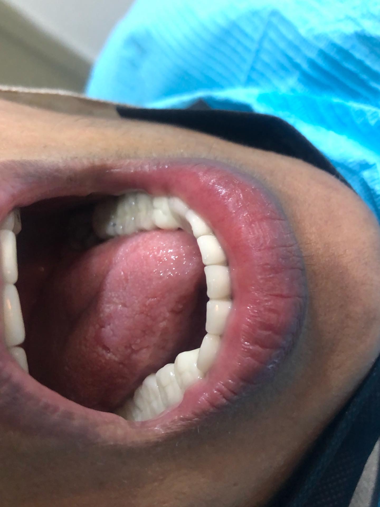
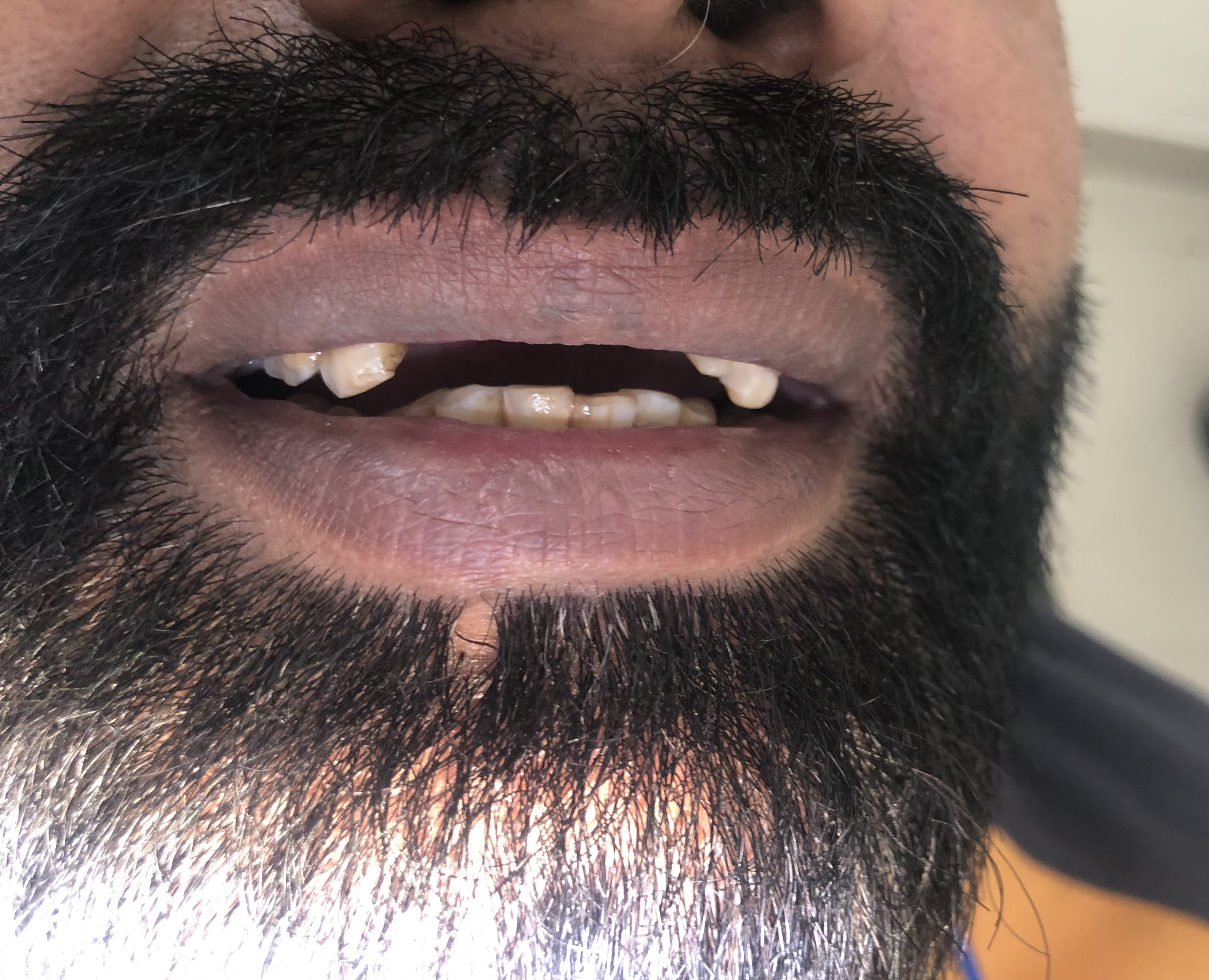
The first thing we saw on examination was the prosthesis itself — a full-mouth fixed bridge that had been placed when Ms. Abida was in tenth grade, more than two decades earlier. The margins had broken down in several places. There were visible black lines at the cervical edge of multiple units, and the metal substructure was beginning to show through in two locations. The original bridge had served her — at one level — for an enormously long time. It had also been hiding the slow accumulation of problems underneath it, and the time had come for those problems to be addressed.
Multiple teeth requiring endodontic therapy
When the old bridge came off, several of the abutment teeth showed clear signs of pulpal compromise. The original clinician, more than twenty years earlier, had prepared multiple teeth for full-coverage restorations without first performing root canal therapy where the depth of preparation made that necessary. That is a clinical choice that was sometimes made in that era. The downstream effect was years of low-grade pulpal inflammation that the patient had not consciously felt, because the cemented bridge had been holding everything still.
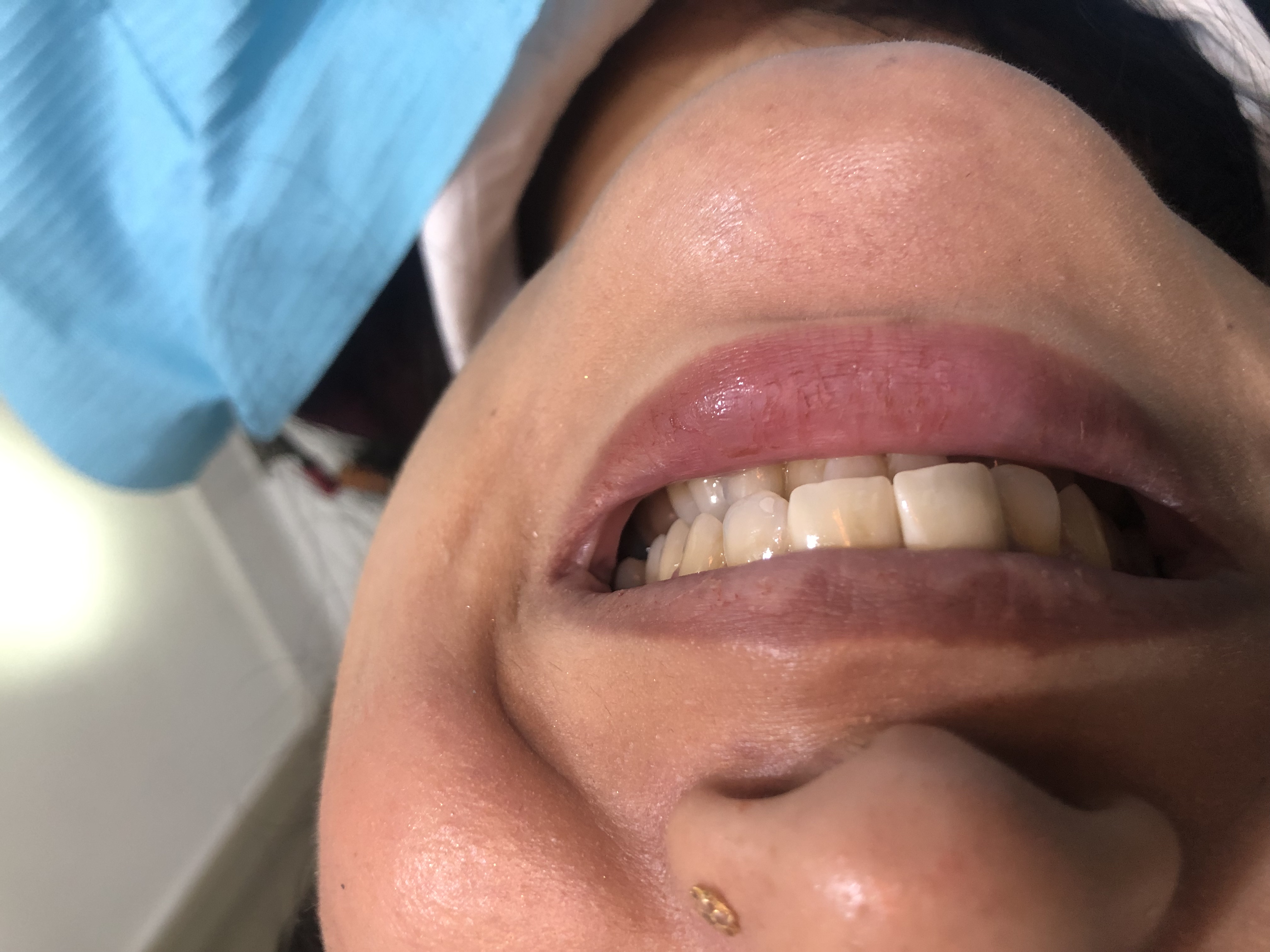
Gingival hyperplasia — the gum had overgrown
The gum tissue around the cervical margins of the old bridge was visibly enlarged. When we probed it, the tissue was spongy, bled on light contact, and had developed several false pockets up to 6 mm. This is a pattern we call gingival hyperplasia — a chronic inflammatory overgrowth of the gum, in this case driven by years of plaque retention at the margins of a bridge whose fit had degraded. It looks alarming, but it is largely reversible once the irritant is removed.
No catastrophic bone loss — the case was rescue-able
The two periapical X-ray sets and the panoramic radiograph we took at the first visit showed something quietly important: the alveolar bone supporting the abutment teeth was, in most areas, intact. There was localised crestal bone loss in two zones where the bridge margins had broken down most severely, but the bone level was still adequate for new abutment teeth. That single finding made the difference between a re-bridging plan and a plan that would have moved her into implants or a removable prosthesis.
Four phases. Eleven weeks. Nine visits.
Each phase was completed before the next began, with a full re-examination at every transition. The patient never went home with an open margin, an unsealed root canal, or a problem that we had not first discussed with her in writing.
Staged removal of the old bridge
We sectioned the old prosthesis under local anaesthesia and removed it in segments — not all in one piece — so that each abutment tooth could be inspected as the bridge came off. Each segment was photographed, labelled and kept. Some patients want their old bridge returned; we always offer it. We charted every abutment tooth on the day of removal: vital or non-vital, restorable or non-restorable, the depth of any cervical caries underneath, and the integrity of the remaining tooth wall.
Visit 1 · ~ 75 minEndodontic phase — multiple root canals over three weeks
The teeth that needed root canal treatment were treated one quadrant at a time across three weekly visits. We used a magnification loupe to locate and instrument every canal, an apex locator to confirm working length on each, and a calcium-hydroxide intracanal medication between sessions on the teeth that required more than one visit. The final obturation was completed with warm vertical compaction of gutta-percha at the end of the endodontic phase, ready for the restorative build-up.
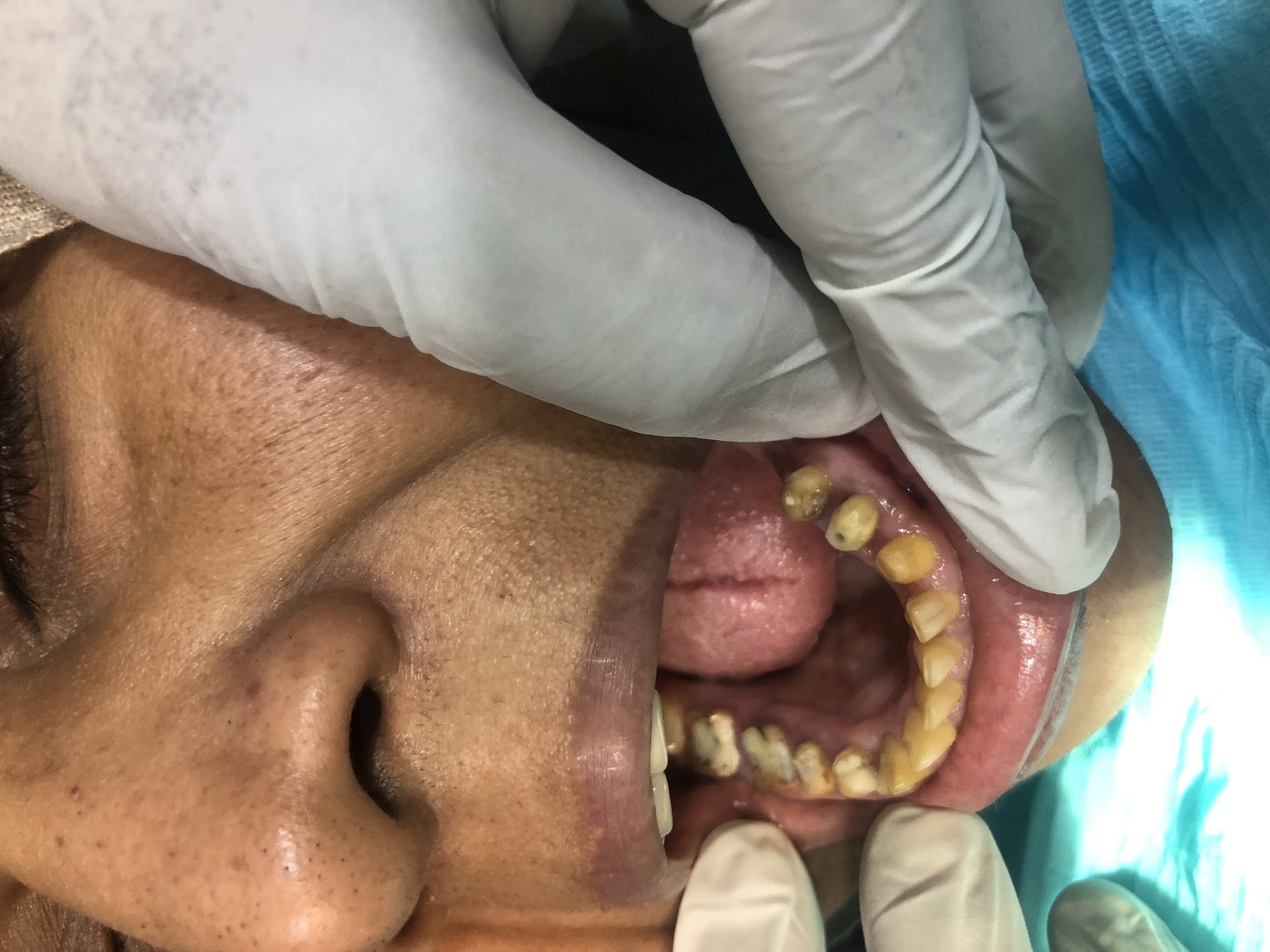
Visits 2-4 · ~ 90 min eachPeriodontal therapy and tissue conditioning
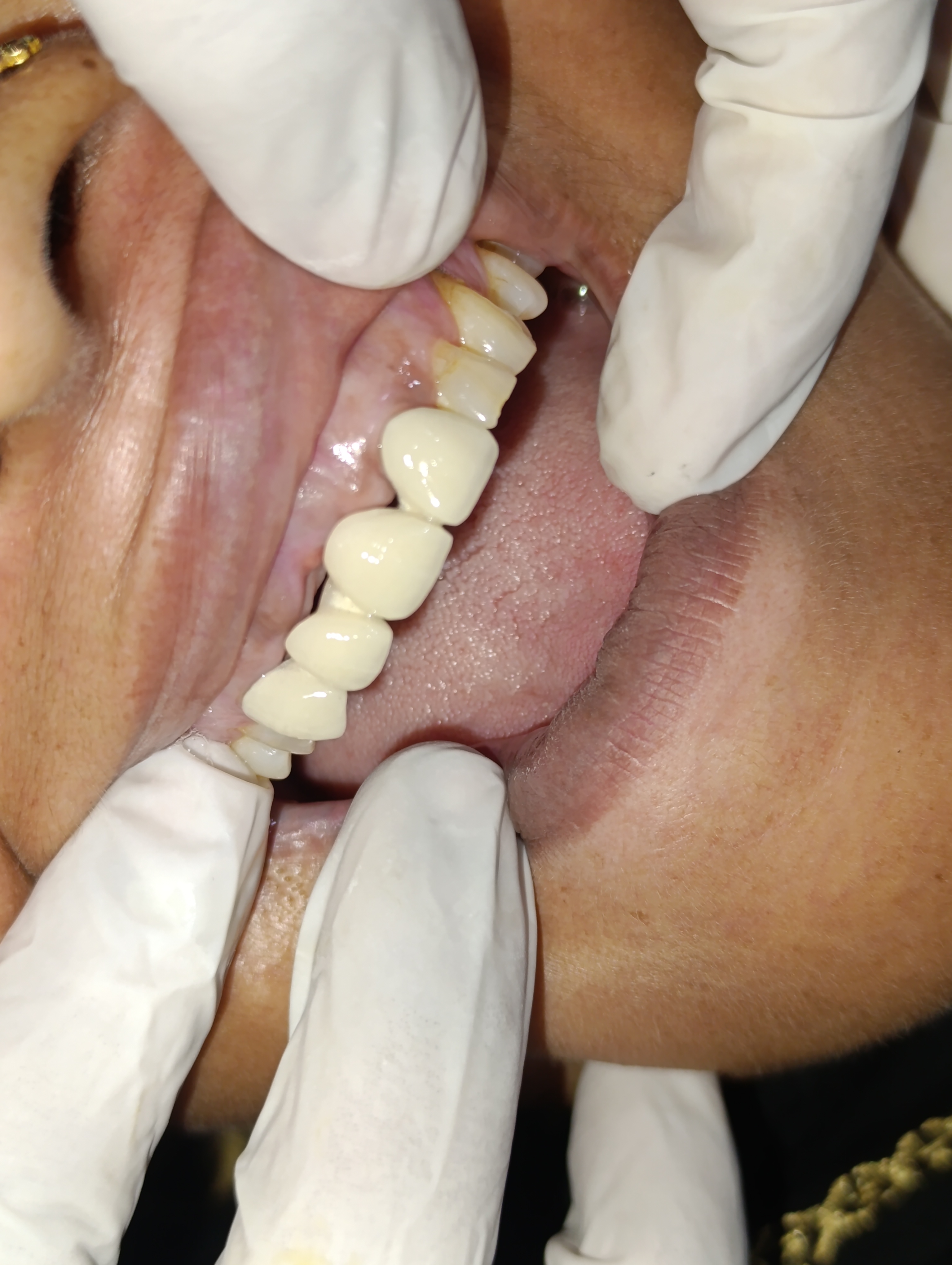
Once the endodontics were finished, we addressed the gum. Full-mouth scaling, root planing in the deeper pockets, and a precise contouring of the hyperplastic tissue using an electrosurgery loop — under local anaesthesia and with a careful, minimal margin. The aim was not to remove the gum but to give it a new, healthier shape that the future bridge margins could meet cleanly. We waited two weeks after this phase for the tissue to mature before any new impressions were taken.
Visit 5 · ~ 60 minNew full-mouth fixed bridge fabrication and delivery
With healthy abutments and a settled gum architecture, we took the master impression for the new bridges. The case was split into two arches handled in sequence — upper first, lower three weeks later. Each arch was tried in at three stages: metal-substructure try-in, biscuit-bake try-in for shade and shape, and final-glaze try-in just before cementation. The new prosthesis was cemented with a self-adhesive resin cement designed for high retention.
Visits 6-9 · across 8 weeks During — endodontic phase complete
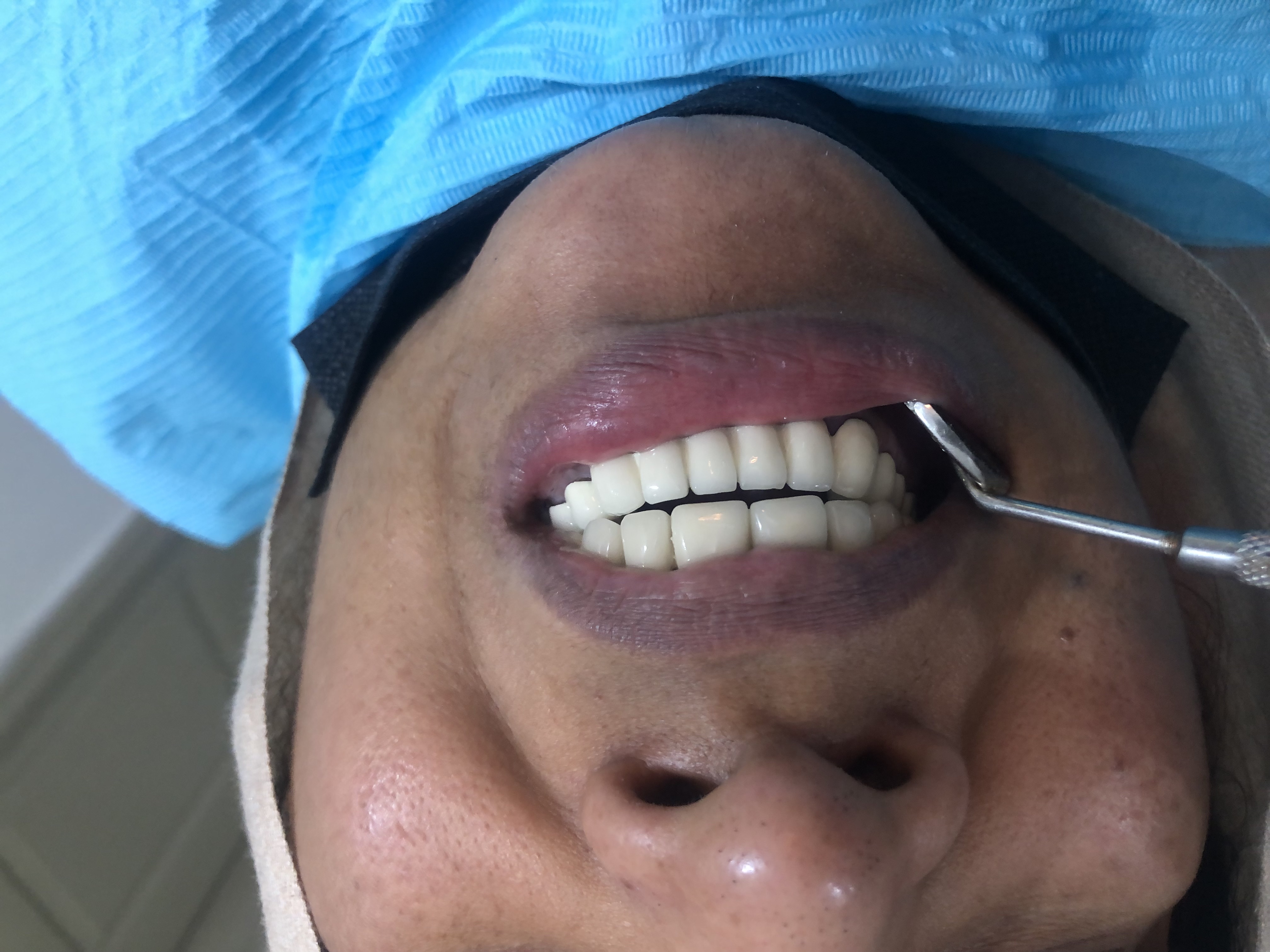 During — periodontal recontouring
Same patient. Twenty-two years apart in dental health.
Drag the divider to compare. The "before" image is from the first consultation visit; the "after" image is from the cementation of the second arch, eleven weeks later.
BeforeAfterWhy a sequenced plan beats a faster one.
Patients asked to undergo a multi-visit rehabilitation reasonably ask whether the same result could have been achieved faster. The honest answer is no — and the reasons are physiological, not administrative.
Endodontically treated teeth need time to confirm symptomatic resolution
After a root canal treatment is completed, the tooth needs a short period of clinical observation — usually one to two weeks — to confirm that there is no residual peri-apical inflammation. Loading that tooth with a permanent bridge before that observation period has passed risks discovering, three months later, that a single abutment needs re-treatment under the new bridge. That is the kind of problem that turns an 11-week case into a 6-month case. Patience now saves rework later.
Gum tissue needs to mature into its new shape before impressions
When hyperplastic gum tissue is recontoured — whether with an electrosurgery loop, a diode laser, or a cold-blade gingivectomy — the new tissue margin takes around 14 days to fully mature. Taking the master impression before that maturation is complete results in a final bridge whose cervical margin is sitting on a moving line of tissue. That is the leading cause of the “black-line-at-the-gum” complaint a year later. We always wait the two weeks.
Each try-in stage is an opportunity to fix a fitting issue before it's permanent
A full-mouth bridge case goes through three try-in stages before cementation: metal-only, biscuit-bake porcelain, and final glaze. Each stage lets us check a different thing — marginal fit, porcelain shade and shape, and final-glaze occlusion. Skipping any of those try-ins risks discovering the problem only after cementation, when the only fix is to break the bridge off and start over. Each try-in costs the patient one short visit. The cost of skipping a try-in is much higher.
Five questions we hear at every full-mouth consult.
These are the worries we heard from Ms. Abida and the worries we hear from most patients who consider a full-mouth rehabilitation. Tap any one to read the long answer.
Could you not have just left the old bridge in place if it was still functional?+
On the surface, yes — Ms. Abida had been chewing with the original bridge for over twenty years, and she came in not because the bridge had failed catastrophically but because she had developed discomfort under it. So a reasonable question to ask is whether we could have intervened more conservatively.
The answer is no, for three reasons. First, the marginal seal of the bridge had broken down in multiple places, and once a cement margin opens, saliva and bacteria are able to get under the crown. From that point, the natural tooth underneath the crown is on a steady trajectory toward decay or pulpal compromise — the only question is how fast. Second, the gum was visibly inflamed and hyperplastic at multiple margins. That inflammation was not going to resolve while the irritant — the old bridge — remained in place. Third, several of the underlying teeth needed root canal therapy that could not be safely performed through the existing prosthesis.
The decision to remove the bridge was therefore a sequencing decision: we removed it because we needed access to do the underlying biological work, not because we wanted the bridge gone for cosmetic reasons. The new prosthesis is the final stage of the plan, not the first.
How long will the new bridge last? Will I be back here in another twenty years?+
A well-made porcelain-fused-to-metal full-mouth bridge, on properly endodontically treated and periodontally healthy abutments, with disciplined home care and six-monthly professional checks, has a published service life of 15 to 20 years. The original bridge that came out of Ms. Abida's mouth had survived 22 years — at the upper end of what we expect — and it could have been longer with regular maintenance.
The single biggest factor is the gum and bone around the abutment teeth. If those stay healthy, the bridge will outlast almost anything else in dentistry. If plaque is allowed to accumulate at the margins, the gum will inflame, recede, and eventually expose root surface. That is the same path that broke down her original bridge.
We will see her every six months for the rest of her time at this clinic, and we will catch any marginal issue at the point where a small intervention is enough. With that schedule kept, twenty years is reasonable.
Why did this case need so many visits? Could it not have been done faster?+
A full-mouth rehabilitation that involves removing an old bridge, performing multiple root canals, addressing gingival hyperplasia, and then fabricating new prosthetics is genuinely a multi-visit case. Compressing it carries real risk.
Specifically, the gum tissue needs time to recover and settle after periodontal therapy before any impression is taken — about two weeks of healing is the minimum. Each root canal treatment needs to be sealed and given a period to confirm there is no residual symptomatic flare. The metal substructure of the new bridge needs to be tried in before the porcelain is added, the porcelain needs to be tried in before the glaze, and the glaze needs to be tried in before cementation. Each of those try-in stages is the patient's opportunity to flag anything that does not look right.
Ms. Abida's case took nine visits across approximately eleven weeks. That is the right pace for a case of this complexity. A six-week version of the same case would have been a higher-risk version. We do not compress for the sake of compression.
Will I be in pain during this process?+
No — almost never. The case is performed under local anaesthesia at every stage where the patient could otherwise feel discomfort. The bridge removal, the root canal treatments, the periodontal therapy, and the new bridge preparation are all done with appropriate buccal and palatal infiltrations or block anaesthesia, with the patient comfortable throughout.
There is a typical pattern of mild soreness for the first 24 to 48 hours after each visit — particularly after the periodontal-therapy visit, when the gum has been recontoured. We prescribed Ms. Abida ibuprofen 400 mg three times a day for two days after that visit, and a chlorhexidine mouthwash twice daily for ten days. She did not require anything stronger at any point.
Once the new bridge is cemented, there is no ongoing discomfort. The cement-line is sealed, the gum is healthy, and the new prosthesis transmits chewing forces in a way that the old, failing bridge had stopped doing.
What if one of the new abutment teeth fails in the future?+
The honest answer is that — over a 15-to-20-year service life — there is a real possibility that one of the abutment teeth will eventually develop a complication. Either a marginal caries lesion that requires re-doing one segment of the bridge, or a late peri-apical lesion on a root-canal-treated abutment that requires a re-treatment.
The bridge has been deliberately designed in two sectional units per arch — not a single continuous unit — so that if one section ever needs revising, only that section comes off and only that section is re-fabricated. The other sections remain in place undisturbed. That design choice now is the reason the eventual maintenance, whenever it is needed, will be smaller in scope.
We discussed this honestly with Ms. Abida at consent stage. She accepted that no dental prosthesis is permanent, and the goal of this rehabilitation is not a forever fix but a high-quality 15-to-20-year fix with a maintenance plan attached.
The follow-up visits.
A full-mouth rehabilitation is finished only after a full six-monthly recall cycle confirms the new bridges are settling well and the gum is responding. Here is how Ms. Abida's follow-up went.
Ms. Abida came in for a thirty-minute review one week after final cementation. We checked the occlusion under articulating paper, polished a single high spot on the lower-left posterior unit, and re-checked the gum at all bridge margins. No bleeding on probing at any site. She reported being able to chew on both sides for the first time in years.
The gum had fully matured. The slight inflammation that had remained at two anterior sites at the one-week visit had resolved completely. Photographs were re-taken so we could compare against the pre-treatment series. She had been flossing under all six pontics every night with super-floss.
Six months out, all bridge margins were sealed, gum probing depths were under 3 mm everywhere, and bleeding on probing was negative at every site. A routine professional clean was performed at the same visit. She has booked the twelve-month review for early 2026.
Dr. Mian Momin Ahmad
“Cases like Ms. Abida's are about sequencing. The technical dentistry is not the hard part — any competent prosthodontist can prepare a tooth or place a crown. The hard part is knowing what order to do things in, and resisting the temptation to compress the timeline to please the patient or finish the case in fewer visits. The old bridge had lasted twenty-two years. The new bridge needs to last twenty more. That kind of longevity is built phase by phase.”
Six habits that protect a full-mouth bridge.
The longevity of a full-mouth rehabilitation depends almost entirely on what the patient does at home between visits. These six habits are what we asked Ms. Abida to commit to.
Floss under the bridge every single night
A fixed bridge has a section called a pontic — the tooth-shaped piece that sits in the gap where a natural tooth used to be. Plaque accumulates under that pontic in a way that ordinary brushing cannot reach. A length of super-floss or a floss-threader, slid under each pontic once a day, is the single most important habit a bridge patient can adopt. Ten seconds per pontic, every night. It is the reason a bridge lasts twenty years instead of eight.
Use a soft brush — and an interdental brush at the abutments
A soft-bristled toothbrush is essential, because the gum at the cervical margin of every abutment crown is the area most vulnerable to recession. We also sized Ms. Abida up for a 0.5 mm interdental brush head, to gently disturb plaque at the embrasures between adjacent crowns. Used once a day, after flossing under the pontics, it covers the only sites a normal toothbrush cannot.
Six-monthly recall is non-negotiable, not optional
Ms. Abida's previous bridge lasted as long as it did partly because of her own home care, but it had never been professionally maintained on a schedule. The new bridges will be checked every six months for marginal integrity, cement washout, gum response and any signs of cervical caries developing at any of the abutments. Twenty minutes per recall, no charge for the check itself.
Tell us about any new tenderness immediately
When a bridge abutment develops a problem, the first sign is almost always a mild tenderness on biting — long before any swelling appears. Patients who learn to flag that single early sign, rather than waiting to see if it passes, end up with bridges that get a small intervention at the right time instead of a big intervention later. We asked Ms. Abida to message us on WhatsApp the day she felt anything different.
Limit very hard or very sticky foods on the front bridge
Modern porcelain-fused-to-metal bridges are strong, but they have an upper limit. We asked Ms. Abida not to bite directly into hard naan crust or chicken bones with the upper front bridge, and not to chew large pieces of toffee or jelly sweets on the bridge units. Slicing food and chewing with the posterior bridge units, which are designed for occlusal loading, is the right approach.
Treat dry mouth — if it ever becomes an issue
A dry mouth — from medication, dehydration, or age — accelerates marginal breakdown around every bridge unit, because saliva is the natural buffer that protects the cement-tooth junction. If Ms. Abida ever develops dry mouth for any reason, we asked her to flag it at the next recall so we could fit her with a higher-frequency check and consider a chlorhexidine or fluoride mouthwash regimen.
A bridge has a service life. Knowing when to retire it is the patient's call — not its.
Many patients with bridges that are 15, 18, 20 years old assume that “if it's still in my mouth it must still be working.” That is rarely true. The bridge will keep functioning, in a degraded way, long after its margins have broken down and the gum has started to inflame — because the body is remarkably tolerant. The patient often does not feel the slow change. They feel only the eventual, undeniable change, by which point the case is more complex.
Ms. Abida came in at year 22. The case was still rescue-able with a new bridge. Another five years and we might have been having a very different conversation about implants. The earlier the conversation, the smaller the eventual intervention.
More on bridges and the conditions behind them.
Three more crown and bridge patients.
Every case in this archive is a real Odonto patient with their written consent. Names are accurate where the patient was happy to share them, ages are real, and every photograph was taken in our Engineers Town clinic.
Carrying an old bridge you're no longer sure about?
The first 15 minutes are free. We will examine your bridge, take any X-rays needed, and put a written opinion in your hand — whether or not it needs replacing today. There is no pressure to book the work the same day.