
Dental X Ray in Lahore for tooth pain, OPG scan and treatment planning.
Dental X ray imaging helps the dentist see what is hidden under enamel, gums and bone. At Odonto, radiographs support diagnosis for tooth pain, cavities, root canals, implants, braces, wisdom teeth and surgical planning.
- Tooth X ray for pain, cavities, infection, abscess and root canal planning.
- OPG style assessment for wisdom teeth, jawbone, missing teeth and surgery planning.
- Radiographs used with clinical testing, not as a replacement for examination.
- Digital imaging helps reduce retakes and makes patient explanation clearer.
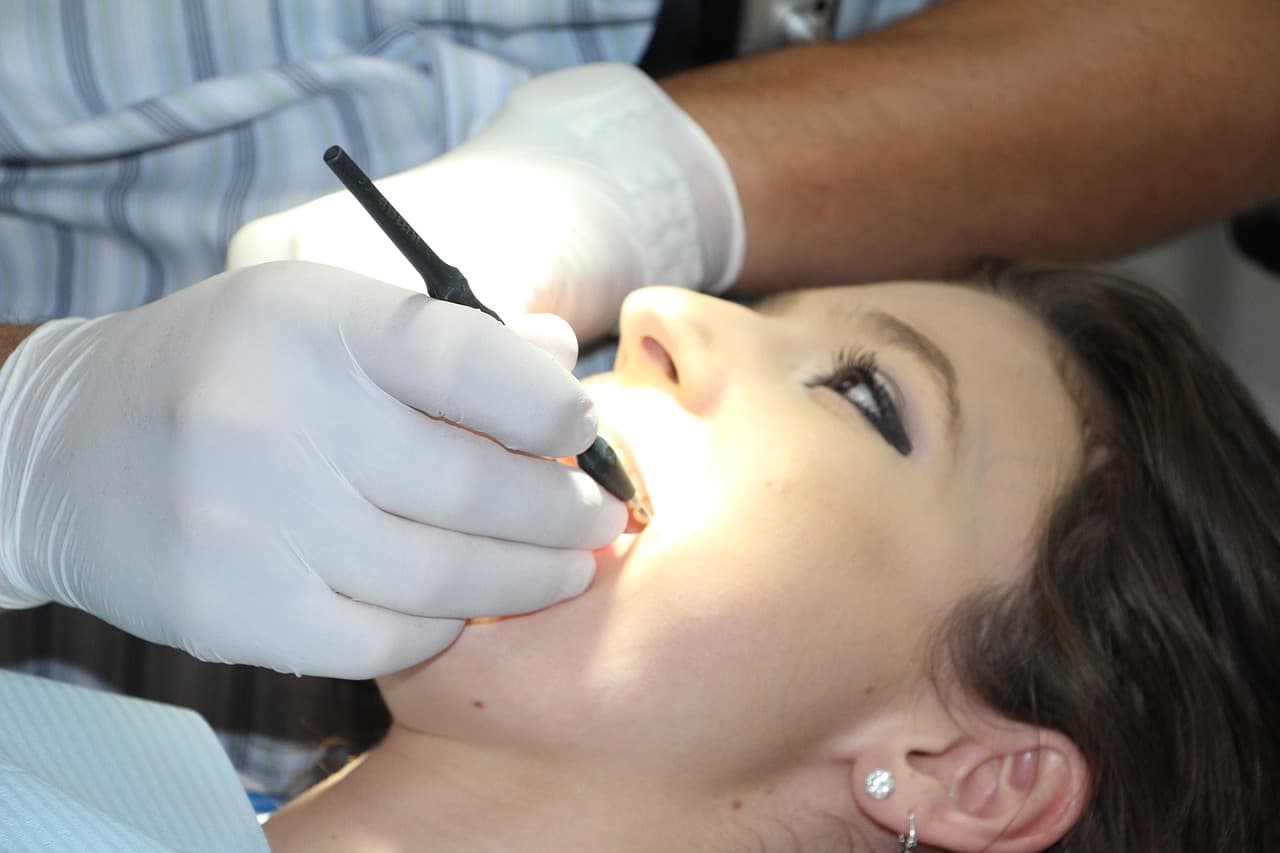
A radiograph helps explain pain that cannot be seen by eye.
Many dental problems begin under the surface. A tooth may look normal but still have deep decay, root infection, bone loss, impacted wisdom tooth pressure or an old filling leaking underneath.
Dental radiography gives the dentist a view of tooth roots, bone level, hidden cavities, infection shadows, tooth position and previous dental work. It is especially useful before root canal treatment, extraction, implant placement and orthodontic planning.
Different images answer different questions. A small periapical image may show one painful tooth in detail. A bitewing can help find hidden cavities between teeth. An OPG gives a broad jaw view for wisdom teeth, missing teeth and overall planning.
X rays should be selected based on need. The goal is not to take every image possible, but to take the right image that changes diagnosis or treatment planning.
Hidden diagnosis
Radiographs help find decay, infection, bone loss and root shape that cannot be judged visually.
Safety minded use
Images are taken when clinically useful and with sensible positioning and exposure control.
Clear explanation
Digital images make it easier to show patients why a treatment is recommended.
Signs you should not ignore.
These symptoms do not always mean surgery or specialist treatment is needed, but they do deserve a proper examination before the problem becomes harder to manage.
Tooth pain
A radiograph may reveal deep decay, infection around the root or a hidden crack related problem.
Swelling or abscess
Images help locate the source tooth and the area of infection.
Wisdom tooth pressure
A jaw view can show angle, depth and relation to nearby teeth.
Before root canal
Root shape, infection and working area are assessed before and during treatment.
Before implant
Bone height, missing spaces and nearby structures must be reviewed before implant planning.
Before braces
Orthodontic records help assess roots, missing teeth, jaw relationship and eruption patterns.
The image should answer a clinical question.
A dental X ray is most useful when the dentist already has a question: is there deep decay, is the root infected, where is the wisdom tooth, is there enough bone, or why does this tooth hurt when the mouth looks normal?
That is why imaging is paired with examination. The radiograph gives evidence, but the diagnosis still depends on symptoms, testing, gum findings and the condition of the tooth.
Care is planned around the diagnosis.
The service list below is here to help you understand the options. At the visit, the plan is narrowed down after examination, radiographs and a clear discussion of benefits, risks and recovery.
Tooth X ray for pain
Used to assess deep cavities, infection, root changes and painful teeth.
OPG X ray discussion
A broad view for wisdom teeth, missing teeth, jawbone and overall treatment planning when indicated.
Root canal radiographs
Images help confirm tooth anatomy, infection and treatment progress.
Implant scan planning
Bone support and nearby structures are assessed before implant recommendations.
Orthodontic imaging
Radiographs support bite, eruption and root assessment before braces or aligners.
Bitewing style cavity check
Images can help detect hidden decay between back teeth where visual exam is limited.
How treatment is planned.
Good treatment starts with the decision before the procedure. The steps stay simple for patients, but the clinical checks behind them are careful.
Clinical question
We identify what the image needs to answer before taking it.
Positioning
The patient is positioned carefully so the area of concern is captured clearly.
Digital image
The radiograph is taken and checked for diagnostic clarity.
Explanation
Findings are explained with the treatment options they affect.
Small tooth image or wider jaw scan?
Different radiographs are useful for different clinical questions.
A small tooth image may suit when
- One tooth is painful or sensitive.
- A root canal, crown or filling is being planned.
- A deep cavity or root infection needs review.
- The dentist needs detail around one or two teeth.
A wider image may suit when
- Wisdom teeth or multiple missing teeth need assessment.
- Jawbone, eruption pattern or overall dentition must be reviewed.
- Implants, braces or oral surgery are being planned.
- The concern involves both sides or several areas.
Dental radiography questions patients ask most.
Good imaging is selective, explained and connected to a treatment decision.
Are dental X rays safe?
Modern dental imaging uses low exposure, and images are taken only when the diagnostic benefit is clear.
Pregnancy and imaging
Tell the dentist if you are pregnant or may be pregnant so the team can decide timing and precautions.
Kids dental images
Children images are chosen carefully based on cavities, eruption, pain or trauma needs.
What a cyst may look like
A cyst or infection can appear as a dark area around bone or roots, but diagnosis needs professional interpretation.

Dr. Mian Momin Ahmad
Consultant Oral Physician and Dental Surgeon
Dr. Mian Momin Ahmad is a general dentist, consultant oral physician and dental surgeon at Odonto.
- Plans restorative, preventive and diagnostic dental care around tooth, gum and bite findings.
- Supports patients with treatment choices for fillings, crowns, endodontic care and tooth replacement planning.
- Focuses on clear explanations, clinical documentation and practical maintenance advice after treatment.
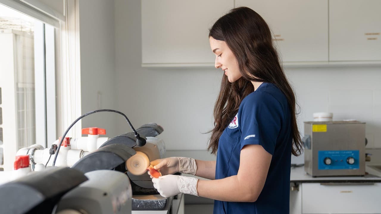
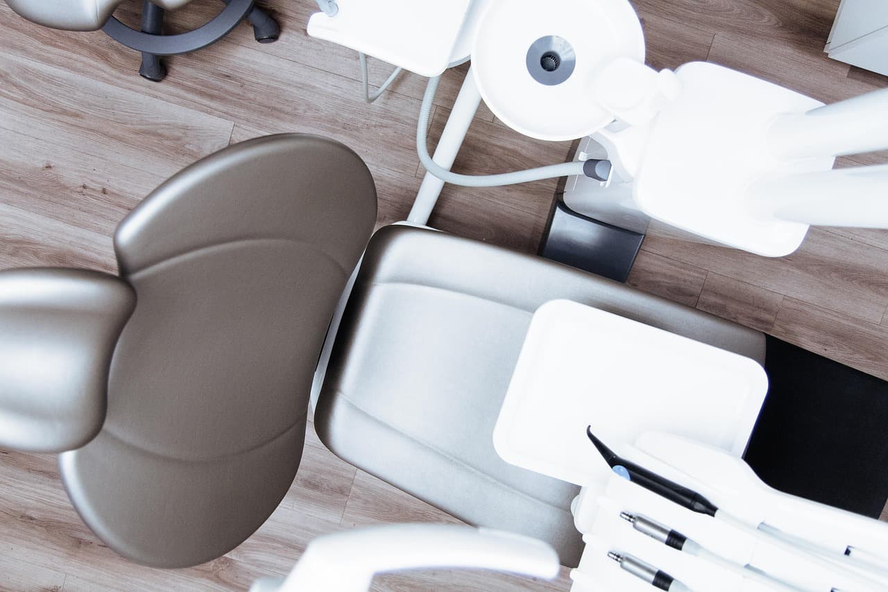
Recent care at Odonto
A look at the clinic environment, treatment planning and service related care. Treatment photographs are shared only where consent is available, and results vary by case.
Want to see more clinic updates and patient education?Follow Odonto on social channels or message us for the most relevant examples before your visit.
Book an appointmentCommon patient questions.
These answers are a starting point. Your exact plan depends on examination, radiographs, medical history and the condition of the tooth, gum, jaw or bite.
What is a dental X ray?
How much is a dental X ray?
What does a cyst look like on a dental X ray?
Do I need an X ray before root canal treatment?
Do I need an X ray before a dental implant?
Are dental X rays safe for children?
Can an X ray show all cavities?
What is an OPG X ray?
Book at Odonto on Main Defence Road.
Share your symptoms on WhatsApp and the team will guide you on appointment timing.
Book at Odonto on Main Defence Road.
Odonto Dental and Aesthetic Clinic is located on Main Defence Road, Block A1, Engineers Town. If you are in acute pain, message first so the team can guide you on timing, pain medicine precautions and whether swelling needs urgent attention.
Bring any old radiographs, prescriptions, crown records or previous treatment notes if you have them. If you do not, that is fine; we can start with a fresh examination and guide the next step after diagnosis.
Phone
+92 320 6373744Hours
Open daily, 11 AM to 10 PM
Address
Plot # 7 Shop # 2, Main Defence Rd, Block A1, Engineers Town, Lahore
Booking
Confirm slot on WhatsAppRelated dental pages.
These internal pages help patients compare nearby treatment options before booking a consultation.
Root canal treatment
Radiographs guide diagnosis and canal planning.
RelatedDental implants
Imaging helps assess bone before implants.
RelatedTooth extraction
X rays help plan tooth removal safely.
RelatedOral surgery
Surgical planning often needs radiographs.
RelatedDental braces
Orthodontic records support bite planning.
RelatedPediatric dentist
Children may need imaging for pain, trauma or eruption.